Home
/ Digestive System Labeled - Associate Degree Nursing Physiology Review : Cardiovascular system and digestive system digestive sistem esophagus stomach organ of the human body anatomy esophagus organs of the digestive system digestive system labeled digestive system illustration digestive system label human body anatomy with labels.
Digestive System Labeled - Associate Degree Nursing Physiology Review : Cardiovascular system and digestive system digestive sistem esophagus stomach organ of the human body anatomy esophagus organs of the digestive system digestive system labeled digestive system illustration digestive system label human body anatomy with labels.
Digestive System Labeled - Associate Degree Nursing Physiology Review : Cardiovascular system and digestive system digestive sistem esophagus stomach organ of the human body anatomy esophagus organs of the digestive system digestive system labeled digestive system illustration digestive system label human body anatomy with labels.. The visceral peritoneum includes multiple large folds that envelope various abdominal organs, holding them to the dorsal surface of the body wall. Food's journey through the digestive system stop 1: Mechanical digestion is the physical breakdown of large pieces of food into smaller pieces. See full list on innerbody.com See full list on courses.lumenlearning.com
See full list on courses.lumenlearning.com The digestive system is made up of the gastrointestinal tract—also called the gi tract or digestive tract—and the liver, pancreas, and gallbladder. The pharynx is responsible for the passing of masses of chewed food from the mouth to the esophagus. The stomach also contains hydrochloric acid and digestive enzymes that continue the digestion of food that began in the mouth. Learn vocabulary, terms, and more with flashcards, games, and other study tools.
Human Digestive System Wikipedia from upload.wikimedia.org Movement of food through the gi tract mucosa: In addition, the mucosa has a thin, smooth muscle layer, called the muscularis mucosa (not to be confused with the muscularis layer, described below). The five major peritoneal folds are described in table 2. This mode of digestion begins with the chewing of food by the teeth and is continued through the muscular mixing of food by the stomach and intestines. Finally, bile is used to emulsify large masses of lipids into tiny globules for easy digestion. Notice that the epithelium is in di. Note that during fetal development, certain digestive structures,. The alimentary canal forms a continuous tube that is open to the outside environment at both ends.
Bile produced by the liver is also used to mechanically break fats into smaller globules.
By the time food has left the duodenum, it has been reduced to its chemical building blocksfatty acids, amino acids, monosaccharides, and nucleotides. The liver is a roughly triangular accessory organ of the digestive system located to the right of the stomach, just inferior to the diaphragm and superior to the small intestine. Because the pharynx serves two different functions, it contains a flap of tissue known as the epiglottis that acts as a switch to route food to the esophagus and air to the larynx. The six primary processes of the digestive system include: Accessory digestive organs comprise the second group and are critical for orchestrating the breakdown of food and the assimilation of its nutrients into the body. Throughout its length, the alimentary tract is composed of the same four tissue layers; Layer of dense connective tissue in the alimentary canal wall that binds the overlying mucosa to the underlying muscularis submucosal plexus: The submucosal plexus(plexus of meissner) lies in. It is composed of two different regions: The blood vessels serving the digestive system have two functions. Mucus serves as a protective barrier and lubricant inside of the gi tract. It is located just inferior to the stomach and takes up most of the space in the abdominal cavity. As soon as food enters the mouth, it is detected by receptors that send impulses along the sensory neurons of cranial nerves.
The entire small intestine is coiled like a hose and the inside surface is full of many ridges and folds. While food is being mechanically digested it is also being chemically digested as larger and more complex molecules are being broken down into smaller molecules that are easier to absorb. See full list on courses.lumenlearning.com Without these nerves, not only would your food be without taste, but you would also be unable to feel either the food or the structures of your mouth, and you would be unable to avoid biting yourself as you chew, an action enabled by the motor branches of cranial nerves. See full list on courses.lumenlearning.com
File Digestive System Diagram En Svg Wikimedia Commons from upload.wikimedia.org Small blood and lymphatic vessels in the intestinal wall pick up the molecules and carry them to the rest of the body. The large intestine is a long, thick tube about 2.5 inches in diameter and about 5 feet long. The blood vessels serving the digestive system have two functions. The liver is a roughly triangular accessory organ of the digestive system located to the right of the stomach, just inferior to the diaphragm and superior to the small intestine. The organs of the alimentary canal are the mouth, pharynx, esophagus, stomach, small intestine, and large intestine. See human digestive system labeled stock video clips. Outermost layer of the alimentary canal wall present in regions within the abdominal cavity submucosa: The final function of the digestive system is the excretion of waste in a process known as defecation.
The first function of the digestive system is ingestion, or the intake of food.
In the stomach and intestines, it is a simple columnar epithelium. It is one among the few important topics, which are repetitively asked in the board examinations. More images for digestive system labeled » The function of this sphincter is to close of the end of the esophagus and trap food in the stomach. Most absorption takes place in the walls of the small intestine, which are densely folded to maximize the surface area in contact with digested food. It uses rhythmic muscle movements (called peristalsis) to force food from the throat into the stomach. Absorption begins in the stomach with simple molecules like water and alcohol being absorbed directly into the bloodstream. Mechanical digestion is the physical breakdown of large pieces of food into smaller pieces. Notice that the epithelium is in di. A few milliliters of watery fluid act as a lubricant to minimize friction between the serosal surfaces of the peritoneum. The small intestine is a long, thin tube about 1 inch in diameter and about 10 feet long that is part of the lower gastrointestinal tract. These enteric neurons are grouped into two plexuses. The organs of the alimentary canal are the mouth, pharynx, esophagus, stomach, small intestine, and large intestine.
The stomach is a muscular sac that is located on the left side of the abdominal cavity, just inferior to the diaphragm. Intrinsic innervation of much of the alimentary canal is provided by the enteric nervous system, which runs from the esophagus to the anus, and contains approximately 100 million motor, sensory, and interneurons (unique to this system compared to all other parts of the peripheral nervous system). It is located just inferior to the stomach and takes up most of the space in the abdominal cavity. See full list on courses.lumenlearning.com At the inferior end of the esophagus is a muscular ring called the lower esophageal sphincter or cardiac sphincter.
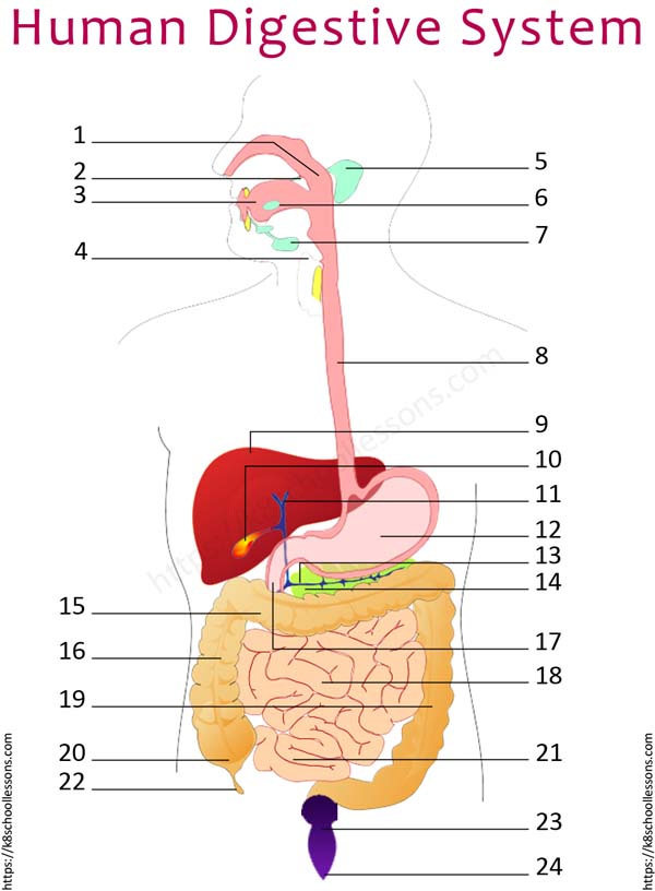
Label Human Digestive System Quiz 1 Digestive System Worksheets from k8schoollessons.com (plexus of auerbach) major nerve supply to alimentary canal wall; This major organ acts as a storage tank for food so that the body has time to digest large meals properly. Bile produced by the liver is also used to mechanically break fats into smaller globules. The pancreas is a large gland located just inferior and posterior to the stomach. The esophagus is a muscular tube connecting the pharynx to the stomach that is part of the upper gastrointestinal tract. See full list on innerbody.com While food is being mechanically digested it is also being chemically digested as larger and more complex molecules are being broken down into smaller molecules that are easier to absorb. The liver weighs about 3 pounds and is the second largest organ in the body.
The parietal peritoneum, which lines the abdominal wall, and the visceral peritoneum, which envelopes the abdominal organs.
It is located just inferior to the stomach and takes up most of the space in the abdominal cavity. The blood vessels serving the digestive system have two functions. Accessory digestive organs comprise the second group and are critical for orchestrating the breakdown of food and the assimilation of its nutrients into the body. See full list on courses.lumenlearning.com In addition, the mucosa has a thin, smooth muscle layer, called the muscularis mucosa (not to be confused with the muscularis layer, described below). Note that during fetal development, certain digestive structures,. See full list on innerbody.com Outermost layer of the alimentary canal wall present in regions within the abdominal cavity submucosa: Accessory digestive organs, despite their name, are critical to the function of the digestive system. The organs of the alimentary canal are the mouth, pharynx, esophagus, stomach, small intestine, and large intestine. The liver weighs about 3 pounds and is the second largest organ in the body. It is one among the few important topics, which are repetitively asked in the board examinations. The gallbladder is used to store and recycle excess bile from the small intestine so that it can be reused for the digestion of subsequent meals.
